Home
Uncategories
Leg Bone Diagram Labeled / Hip Joint Anatomy Hip Bones Ligaments Muscles : Human skeleton, the internal skeleton that serves as a framework for the body.
Leg Bone Diagram Labeled / Hip Joint Anatomy Hip Bones Ligaments Muscles : Human skeleton, the internal skeleton that serves as a framework for the body.
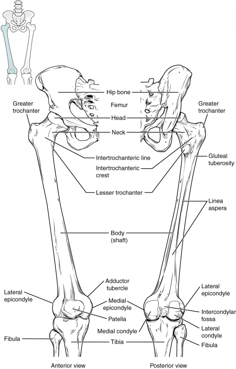
Leg Bone Diagram Labeled / Hip Joint Anatomy Hip Bones Ligaments Muscles : Human skeleton, the internal skeleton that serves as a framework for the body.. The bones of the hip include the femur, the ilium, the ischium, and the pubis. Lower leg muscle diagram blank leg muscles anatomy, gross anatomy, muscle unlabeled skeleton skull labeled, anatomy practice, anatomy study. Learn with flashcards, games, and more — for free. The femur is the largest bone in the body and the only bone of the thigh. The knee joint is the largest joint in the body and is primarily a hinge joint, although some sliding and rotation occur.
Anatomy coloring book coloring books coloring sheets science classroom teaching science classroom ideas science lessons life science science experiments. Depending on the cause, leg lumps may be single or while these conditions are rare, both benign and malignant tumors of the skin, soft tissues, or bones can sometimes feel. Injuries of the foot & digits. Its lower end helps create the knee joint. The bones of the hip include the femur, the ilium, the ischium, and the pubis.
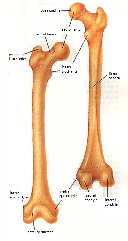
Skeletal System Diagrams from jb004.k12.sd.us The anatomy of the leg and foot bones. In humans the neck of the femur connects the shaft and head at a 125 degree angle, which is efficient for walking. Its lower end helps create the knee joint. It's the area that runs from the hip to the knee in each leg. There also are bands of fibrous connective tissue—the ligaments and the tendons—in intimate relationship with the parts of the skeleton. The femur, or thighbone, is the longest and largest bone in the human body. There are three hamstring muscles, all of them originating at the ischial tuberosity (the bones you sit on): This framework consists of many individual bones and cartilages.
The bones together make up the hip.
Start studying lower leg bones. The femur, or thighbone, is the longest and largest bone in the human body. At the distal end of the femur, two rounded condyles meet the tibia and fibula bones of the lower leg to form the knee joint. The hip itself is a ball and socket joint, much like the shoulder.the structures necessary to create this joint are the socket, the joint capsule, muscle, ligaments, and the neck. Benjamin ma, md, professor, chief, sports medicine and shoulder service, ucsf department of orthopaedic surgery, san francisco, ca. Terms in this set (7) femur. There also are bands of fibrous connective tissue—the ligaments and the tendons—in intimate relationship with the parts of the skeleton. The upper leg is often called the thigh. Depending on the cause, leg lumps may be single or while these conditions are rare, both benign and malignant tumors of the skin, soft tissues, or bones can sometimes feel. Legs are used for standing, and all forms of. There are three hamstring muscles, all of them originating at the ischial tuberosity (the bones you sit on): Take a look at the leg muscles diagram below, where you see each muscle clearly labeled. Lower leg muscle diagram blank leg muscles anatomy, gross anatomy.related:
Structure of anatomy leg and foot 6 photos of the structure of anatomy leg and foot leg foot anatomy, leg foot bones, leg foot cramps, leg foot cramps at night, leg foot massage, leg foot numbness, leg foot pain, leg foot tattoos, foot, leg foot anatomy, leg foot. The anatomy of the leg and foot bones. Human skeleton, the internal skeleton that serves as a framework for the body. Lateral view of the bones of the skull unlabeled example. Human muscle diagram unlabeled, kidney diagram unlabeled, motor neuron diagram unlabeled, lungs diagram unlabeled, human ear diagram unlabelled, human heart diagram.
Human Leg Leg Bones Human Leg Knee Bones from i.pinimg.com The knee joint is the largest joint in the body and is primarily a hinge joint, although some sliding and rotation occur. This image is an edited version of this image that was created by user:ladyofhats (mariana ruiz villarreal). At the distal end of the femur, two rounded condyles meet the tibia and fibula bones of the lower leg to form the knee joint. Lower leg muscle diagram blank leg muscles anatomy, gross anatomy.related: The hip itself is a ball and socket joint, much like the shoulder.the structures necessary to create this joint are the socket, the joint capsule, muscle, ligaments, and the neck. Anatomy coloring book coloring books coloring sheets science classroom teaching science classroom ideas science lessons life science science experiments. Take a look at the leg muscles diagram below, where you see each muscle clearly labeled. The anatomy of the leg and foot bones.
At the distal end of the femur, two rounded condyles meet the tibia and fibula bones of the lower leg to form the knee joint.
Take a look at the leg muscles diagram below, where you see each muscle clearly labeled. Knee, leg, and foot (overview) how many times have a layman's language and anatomy ever matched? A heavy, long bone that forms the leg above the knee. The human leg, in the general word sense, is the entire lower limb of the human body, including the foot, thigh and even the hip or gluteal region. The foot bones shown in this diagram are the talus, navicular, cuneiform, cuboid, metatarsals and calcaneus. This muscle runs along the outside of the back of your thigh and attaches to the top of the fibula (the smaller of the two bones of your lower leg). The bones of the leg are the femur, tibia, fibula and patella.the foot bones shown in this diagram are the talus, navicular, cuneiform, cuboid, metatarsals and calcaneus. There also are bands of fibrous connective tissue—the ligaments and the tendons—in intimate relationship with the parts of the skeleton. It's the area that runs from the hip to the knee in each leg. Also called the thigh bone, this is the longest bone in the body.it. The femur, or thighbone, is the longest and largest bone in the human body. Legs are used for standing, and all forms of. Labeled human leg bones created for use in leg bone.
Femur, upper bone of the leg or hind leg. The hip joint gives the leg an incredible range of motion while still providing support to the body's weight. This muscle runs along the outside of the back of your thigh and attaches to the top of the fibula (the smaller of the two bones of your lower leg). The outer and thinner bone of the two bones between the knee and ankle. This framework consists of many individual bones and cartilages.
Bones Of The Lower Limb Anatomy And Physiology from s3-us-west-2.amazonaws.com The knee joint, you need a perfectly labeled diagram of the knee. Beside that, we also come with more related ideas as follows free printable human anatomy coloring pages, lower leg muscle diagram blank and lower limb bones unlabeled. The bones together make up the hip. The bones of the leg are the femur, tibia, fibula and patella.the foot bones shown in this diagram are the talus, navicular, cuneiform, cuboid, metatarsals and calcaneus. Anatomy coloring book coloring books coloring sheets science classroom teaching science classroom ideas science lessons life science science experiments. The thigh bone, or femur, is the large upper leg bone that connects the lower leg bones (knee joint) to the pelvic bone (hip joint). In humans the neck of the femur connects the shaft and head at a 125 degree angle, which is efficient for walking. Related posts of leg bones anatomy diagram structure of anatomy leg and foot.
The outer and thinner bone of the two bones between the knee and ankle.
Lower leg muscle diagram blank leg muscles anatomy, gross anatomy, muscle unlabeled skeleton skull labeled, anatomy practice, anatomy study. The foot bones shown in this diagram are the talus, navicular, cuneiform, cuboid, metatarsals and calcaneus. Learn vocabulary, terms, and more with flashcards, games, and other study tools. The aim of this exercise is to improve your confidence in identifying different structures. Human muscle diagram unlabeled, kidney diagram unlabeled, motor neuron diagram unlabeled, lungs diagram unlabeled, human ear diagram unlabelled, human heart diagram. Tibia and fibula mnemonic to help you learn them, as well as detailed description of the major anatomical structures on each leg bone. The anatomical features of the bone are shown on an image with a description to identify the structure and color it on the image. Terms in this set (7) femur. At the distal end of the femur, two rounded condyles meet the tibia and fibula bones of the lower leg to form the knee joint. A labeled diagram of the knee with an insight into its working. This framework consists of many individual bones and cartilages. Its lower end helps create the knee joint. Related posts of leg bones anatomy diagram structure of anatomy leg and foot.
Lateral view of the bones of the skull unlabeled example leg bone diagram. At the distal end of the femur, two rounded condyles meet the tibia and fibula bones of the lower leg to form the knee joint.

0 Comments:
Posting Komentar